By Sara McPherson
Cell division is an important part of what allows us to grow and reproduce. Cell division is also involved in many important processes like cell differentiation, healing from injuries, and fighting infections, all of which help us survive as multicellular organisms. But how do cells go through division to accomplish these tasks? There are two different methods that a cell can go through division, depending on if reproduction is the end goal or not. The two types of division are known as mitosis and meiosis.
Mitosis is cell division that results in two genetically identical cells from the original cell. Mitosis is the primary method of cell division and will occur millions of times in our bodies and allows us to grow from a zygote to a fully grown adult. It is an important aspect of our everyday lives and is a constant occurrence throughout our bodies. Meiosis, however, occurs at select regions of the body and its only use is in reproduction. Cells undergoing meiosis will divide twice, and result in 4 genetically different cells called gametes. Gametes contain exactly half of the genetic information of a typical cell found in the human body. In order for reproduction to occur and for these cells to be considered viable, a female and a male gamete must come together and form a new and genetically unique cell. A big difference between these two types of cellular division is the resulting cells and their genetic components. They both start with diploid cells and meiosis results in 4 haploid cells, while mitosis results in 2 diploid cells.
Humans are composed of diploid cells, which means that each cell has two copies of each set of chromosomes (we get one set from our mother and one set from our father). Mitosis essentially makes an identical copy of every chromosome and then divides the cell so that each one will have the exact same set of chromosomes that the parent cell starts with. Meiosis goes through the same replication process so that the chromosomes double, but there will be two division steps. The first division will result in two diploid cells just like mitosis, but these cells will not be genetically identical (although they will have the proper number of each chromosome). The second division will take these two diploid cells and divide them, so each has one complete set of chromosomes, making all four of these cells haploid. Essentially, a haploid cell will have one set of chromosomes and a diploid cell will have two sets of chromosomes.
It is important that meiosis results in these haploid cells called gametes because two haploid cells will come together and form a diploid cell known as a zygote. In humans, this zygote will go through many mitotic divisions to eventually form an embryo and then a fetus, eventually resulting in a baby.
The following diagram shows a simplified process of how mitosis and meiosis occur. More occurs within these processes than what is indicated in this diagram, however, this diagram shows the end result of these processes and the main differences that occur in each cell.

Although cell division is a daily occurrence in our bodies, that does not mean that it is simple. Each division will go through a series of steps that must be successfully completed to get viable cells. The following sections give a more detailed description of the stages of mitosis and meiosis.
Mitosis
Prophase

At this point, DNA replication has already occurred. During prophase, the chromosomes begin to condense, the nuclear envelope begins to degrade, and mitotic spindles form. These spindles will be able to ‘attach’ to the chromosomes and organize them, eventually pulling them apart later in mitosis.
Metaphase

The chromosomes align at the metaphase plate to prepare for separation, and the chromosomal kinetochores attach to the microtubules that will eventually pull the chromosomes apart.
Anaphase

At this stage, the sister chromatids (duplicated chromosomes) begin to pull apart to opposite sides of the cell and the cell begins to elongate and prepares for cell division.
Telophase

Telophase is the final mitotic stage before the cell separates, therefore normal cellular structures begin to reform. This occurs as the spindle fibres start to break down, two nuclear membranes begin to form, and the chromosomal DNA becomes less condensed.
Meiosis
Prophase I

Chromosomes condense at this stage and will pair up with their matching chromosome (ex. Chromosome 1 pairs up with chromosome 1). At this point, crossing-over events occur. This means that pieces of the paired chromosomes essentially switch with each other. This causes greater genetic diversity, which will be helpful in the case of reproduction.
Metaphase I

During Metaphase I, the pairs of chromosomes align at the metaphase plate with random orientation so that when the chromosomes pull apart, the chromosome that will move to one side will be random. This also allows for further genetic diversity.
Anaphase I

The chromosome pairs begin to separate so that a full chromosome is pulled to each side of the cell.
Telophase I

The chromosomes are fully separated at this stage and reach opposite ends of the cell.
Prophase II

Similar to prophase in mitosis, spindle fibres form and attach to the sister chromatids.
Metaphase II

The chromosomes align at the metaphase plate.
Anaphase II

The sister chromatids are pulled apart by the spindle fibres.
Telophase II

The final meiotic stage occurs with a nuclear membrane forming around each set of chromosomes and the DNA becomes less condensed.
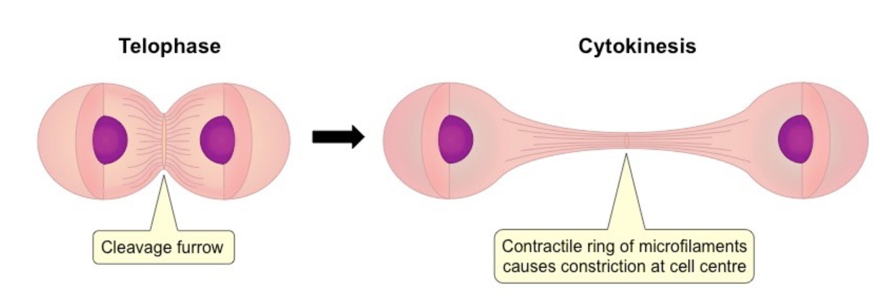
Interphase and Cytokinesis
These stages occur within the life cycle, but may not explicitly be considered part of the mitotic or meiotic cell cycle. Interphase is the cell stage where everything is “normal”; it is the stage that the cell is in when it is not going through mitosis or meiosis. Essentially, a cell in interphase is simply living. Cytokinesis is considered part of cell division but is not always considered part of mitosis or meiosis. The stage of cytokinesis is the cell division stage, where the cell (but not the genetic material) divides. This stage is necessary for mitosis and meiosis to occur, but is often not considered a stage in mitosis or meiosis because all of the cellular materials have already divided. Cytokinesis is also seen as an extension of telophase because of its role in cell division. The following image shows the distinction between telophase and cytokinesis.
Works Cited
- Phases of mitosis | mitosis | biology (article) [Internet]. Khan Academy. Khan Academy; [cited 2022Jun7]. Available from: https://www.khanacademy.org/science/ap-biology/cell-communication-and-cell-cycle/cell-cycle/a/phases-of-mitosis
- Meiosis | cell division | biology (article) [Internet]. Khan Academy. Khan Academy; [cited 2022Jun7]. Available from: https://www.khanacademy.org/science/ap-biology/heredity/meiosis-and-genetic-diversity/a/phases-of-meiosis
- Mitosis and meiosis – comparison chart, video and pictures [Internet]. Diffen. [cited 2022Jun7]. Available from: https://www.diffen.com/difference/Meiosis_vs_Mitosis
- Life Sciences Cyberbridge. [cited 2022Jun7]. Available from: http://cyberbridge.mcb.harvard.edu/mitosis_5.html
- Life Sciences Cyberbridge. [cited 2022Jun7]. Available from: http://cyberbridge.mcb.harvard.edu/mitosis_6.html
- Life Sciences Cyberbridge. [cited 2022Jun7]. Available from: http://cyberbridge.mcb.harvard.edu/mitosis_7.html
- Cytokinesis proteins research tools – creative BioMart [Internet]. Cytokinesis Proteins Research Tools – Creative BioMart – Creative BioMart. [cited 2022Jun7]. Available from: https://www.creativebiomart.net/researcharea-cytokinesis-proteins_923.htm
Sara is a fourth-year biology student at the University of Waterloo, with a particular passion for genetics and anything molecular biology. She is currently working as a co-op student at the BC Cancer Center where she is enjoying studying cancer research and cancer genetics. Outside of her studies, Sara enjoys playing a variety of sports. In the winter you can find her at the rink playing ice hockey and during the summer outside playing soccer.
[/et_pb_text][/et_pb_column][/et_pb_row][/et_pb_section]